You're about to create your best presentation ever

Presentation Background Aorta

Create your presentation by reusing one of our great community templates.

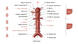
AORTA

Transcript: AORTA Aorta Conditions WHAT IS THE AORTA? Aortic atherosclerosis: Cholesterol plaques build up in the wall of the aorta, posing a risk for stroke. High blood pressure and abnormal cholesterol levels are often responsible. Aortic aneurysm: A weakness in the aorta's wall allows a section to expand like a balloon. Aortic aneurysms are slow to grow, but they can be fatal if they rupture. Aortic dissection: High blood pressure and/or damage to the aorta's wall cause the wall's layers to separate. Aortic dissection can be life-threatening. Aortic regugitation: The aortic valve does not close completely, allowing some blood to flow back into heart with each beat. Autoimmune diseases, Marfan's syndrome, or endocarditis can cause aortic insufficiency. The aorta is the largest artery in the body. Arteries are vessels that carry blood away from the heart. The aorta arises from the left ventricle of the heart, forms an arch, then extends down to the abdomen, where it branches off into two smaller arteries. AORTIC CONDITIONS Aortic stenosis: Narrowing of the aortic valve, which causes the heart to strain to pump blood through. Conclusion Branches of the Aorta Function of the Aorta The aorta carries and distributes oxygen rich blood to all arteries. Most major arteries branch off from the aorta, with the exception of the main pulmonary artery. Ascending Aorta - Extends from the left ventricle of the heart. Aortic Arch - Three major arteries originate from the aortic arch: the brachiocephalic artery, which supplies blood to the brain and head; the left common carotid artery and the left subclavian artery. Descending Aorta - Although the descending aorta is positioned to the left of the body's midline, it gradually descends to directly in front of the vertebral column at the left of the 12th thoracic vertebra. The portion of the descending aorta above the diaphragm is called the thoracic aorta, and gives off branches into the thoracic wall. Below the diaphragm, the descending aorta becomes the abdominal aorta and stems off into branches that reach the abdominal wall and various tissues and organs of the abdomen. Structure of the Aortic Walls The walls of the aorta consist of three layers. They are the tunica adventitia, the tunica media, and the tunica intima. These layers are composed of connective tissue, as well as elastic fibers. These fibers allow the aorta to stretch to prevent over-expansion due to the pressure that is exerted on the walls by blood flow.

aorta

Transcript: The right atrium is one of the four hollow chambers of the interior of the heart. It is located in the upper right corner of the heart superior to the right ventricle. Pulmonary vein Cassandra kyle landiza Student: The pulmonary valve is the semilunar valve of the heart that lies between the right ventricle and the pulmonary artery and has three cusps. Similar to the aortic valve, the pulmonary valve opens in ventricular systole, when the pressure in the right ventricle rises above the pressure in the pulmonary artery. Pulmonary artery left ventricle The left ventricle is one of four chambers of the heart. It is located in the bottom left portion of the heart below the left atrium, separated by the mitral valve. As the heart contracts, blood eventually flows back into the left atrium, and then through the mitral valve, whereupon it next enters the left ventricle. From there, blood is pumped out through the aortic valve into the aortic arch and onward to the rest of the body. Left antrium Pulmonary valve right ventricle The superior vena cava (SVC) is the superior of the two venae cavae, the great venous trunks that return deoxygenated blood from the systemic circulation to the right atrium of the heart. The pulmonary artery carries deoxygenated blood from the heart to the lungs. It is one of the only arteries (other than the umbilical arteries in the fetus) that carries deoxygenated blood. Tricuspid valve Right antrium The mitral valve, also known as the bicuspid valve or left atrioventricular valve is a dual-flap valve in the heart that lies between the left atrium and the left ventricle. The mitral valve and the tricuspid valve are known collectively as the atrioventricular valves because they lie between the atria and the ventricles of the heart and control the flow of blood. Aortic valve The tricuspid valve, or right atrioventricular valve, is on the right dorsal side of the mammalian heart, between the right atrium and the right ventricle. The function of the valve is to prevent back flow of blood into the right atrium. The aorta is the main artery in the human body, originating from the left ventricle of the heart and extending down to the abdomen, where it splits into two smaller arteries The aorta distributes oxygenated blood to all parts of the body through the systemic circulation Mitral valve Parts Of The heart Aorta The pulmonary veins are large blood vessels that receive oxygenated blood from the lungs and drain into the left atrium of the heart. There are four pulmonary veins, two from each lung. The pulmonary veins are among the few veins that carry oxygenated blood. The inferior vena cava is the inferior of the two venae cavae, the large veins that carry deoxygenated blood from the body into the right atrium of the heart. The inferior vena cava carries blood from the lower half of the body whilst the superior vena cava carries blood from the upper half of the body. It is formed by the joining of the right and the left common iliac veins, usually at the level of the fifth lumbar vertebra. The left atrium is one of the four chambers of the heart, located on the left posterior side. Its primary roles are to act as a holding chamber for blood returning from the lungs and to act as a pump to transport blood to other areas of the heart. superior vena cava Science Project Inferior vena cava The right ventricle is the chamber within the heart that is responsible for pumping oxygen-depleted blood to the lungs. The right ventricle is one of the heart's four chambers. It is located in the lower right portion of the heart below the right atrium and opposite the left ventricle. parts of the heart The aortic valve is one of the two semilunar valves of the heart, the other being the pulmonary valve. The heart has four valves and the other two are the mitral and the tricuspid valves. The aortic valve normally has three cusps or leaflets, although in 1–2% of the population it is found to congenitally have two leaflets.[1] It lies between the left ventricle and the aorta. Submitted to: mr. jeffrey espiritu

Aorta

Transcript: Bicuspid Valve any of the tubes forming part of the blood circulation system of the body, carrying in most cases oxygen-depleted blood TOWARD the heart essentially tubes that collapse when their lumens are not filled with blood Veins are less muscular than arteries and are often closer to the skin valve between the left atrium and the left ventricle is an aortic valve that only has two leaflets, instead of three regulates blood flow from the heart into the aorta, the major blood vessel that brings blood to the body Veins one of the heart valves first one that blood encounters as it enters the heart stands between the right atrium and the right ventricle it allows blood to flow only from the atrium into the ventricle Pictures! (: Vena Cava chamber in the heart that is responsible for pumping deoxygended blood to the lungs located in the lower left portion of the heart below the right artium and opposite of the left ventricle As deoxygenated blood flows into the right atrium, it passes through the tricuspid valve and into the right ventricle, which pumps the blood up through the pulmonary valve and through the pulmonary artery to the lungs artey that supplies blood to the heart called the coronary arteries because they encircle the heart in the manner of a crown run along the outside of the heart and have small branches that dive into the heart muscle to bring it blood Intresting Facts about the Circulatory System Aorta Tricuspid Valve vein carrying oxygenated blood from the lungs to the left artium of the heart one of the four vessels that carry aerated blood from the lungs to the left artium of the heart only veins that carry bright-red oxygentated blood The Circulatory System Right Ventricle left lower chamber of the heart, below the left artium receives blood from the left artium and pumps it out under pressure through the aorta to the body separated by the mitral valve as the heart contracts, blood will flow back into the left atrium, and then through the mitral valve, where it next enters the left ventricle Right Atrium Left Ventricle Pulmonary Vein If you were to lay out all of the arteries, capillaries and veins in one adult, end-to-end, they would stretch about 60,000 miles (100,000 kilometers) Capillaries are tiny, averaging about 8 microns (1/3000 inch) in diameter, or about a tenth of the diameter of a human hair An adult human has an average resting heart rate of about 75 beats per minute, the same rate as an adult sheep the ancient Egyptians were cardiocentric — they believed the heart, rather than the brain, was the source of emotions, wisdom and memory, among other things After circulating within the body for about 120 days, a red blood cell will die from aging or damage any of the muscular-walled tubes forming part of the circulation system by which blood (mainly that which has been oxygenated) is carried away from the heart to all parts of the body blood in arteries is usually full of oxygen, the hemoglobin in the red blood cells is oxygenated. The resultant form of hemoglobin (oxyhemoglobin) is what makes the arterial blood look bright red The Human Heart - Main Parts main artery of the body supplies oxygenated blood to the circulatory system in humans, it passes over heart from the left ventricle and runs down in front of the backbone is a tube that is about a foot long and is just about 2 inches long Like all arteries, the aorta's wall has several layers: The intima, the innermost layer, provides a smooth surface for blood to flow across. The media, the middle layer with muscle and elastic fibers, allows the aorta to expand and contract with each heartbeat. The adventitia, the outer layer, provides additional support and structure to the aorta artery that carries blood from the right ventricle of the heart to the lungs for oxygenation this is the last segment of a long journey for circulating blood known as pulmonary trunk, which is relatively short and wide branches off in two directions -- the left & right pulmonary artery Arteries one of the four chambers of the heart hollow structure deoxygenated blood enters the right atrium through the inferior and superior vena cava located in upper right corner above the right ventricle largest vein in the body carries deoxygenated blood into the heart There are two in humans, the inferior vena cava (carrying blood from the lower body) and the superior vena cava (carrying blood from the head, arms, and upper body) Pulmonary Artery Coronary Vessels

Now you can make any subject more engaging and memorable