You're about to create your best presentation ever

Eye Presentation Background

Create your presentation by reusing one of our great community templates.

Eye Presentation

Transcript: Visual Transduction Process By: Dylan Blake What is it? Visual transduction is the process that the eye goes through to be able to see. Simply put, light enters the eye through the hole known as the pupil and is focused by the lens. The lens focuses light on the back of the eye, the retina, where the rods (black and white light detection) and the cones (color detection) are located. From there the message is passed down the photoreceptors and to the ganglion cells which make up the optic nerve, taking the information to where it is processed in the brain. Light is passed over the transparent cornea which roughly begins to focus the light onto the back of the eye known as the retina. It also focuses light into the eye and onto the main focusing part of the eye, the lens. Cornea Cornea The lens is the main component of the eye that deals with focusing the incoming light on the back of the eye on the fovea. Lens Lens The fovea is the place in the retina that holds the most highly concentrated location of photoreceptors in the eye that interprets color and everything around us. The fovea is the locaton of the eye in which light is focused. If the focus point from the lens is before the fovea, then the person will be nearsighted, but if the person's focus point from the lens lies beyond the fovea, they will be farsighted. Fovea Fovea The retina is the location of all of the photoreceptors and cells in an eye. The retina is where the eye absorbs and sends information from the photoreceptors (rods and cones) to the optic nerve and later the brain. Retina Retina As light enters the eye, it will first hit onto the ganglion cells in the retina which is then sent to the bipolar cells and finally the photoreceptors athe the back. From there, impulses are sent in the opposite direction from the rods and cones to the ganglion cells. What happens in the retina? Light interpretation Rods: -About one billion exist in the retina -Detect black and white light -Multiple rods attatched to one neuron (low visual acuity) Cones: -About three million exist only in the fovea -Detect red blue and green light according to the trichromatic theory which is contested by the opponent process theory that states that receptors come in pairs (red/green, blue/yellow, and black/white) -Each cone has its own neuron making for a higher visual acuity. Rods Vs. Cones Photoreceptors The optic nerve is made up of ganglion cells that come from the photoreceptors which receive the light and send impulses to the ganglion cells that are then bunched together to form the optic nerve. The optic nerve runs from the retina to the brain and transmits sensory information to the visual cortex. Optic Nerve Optic Nerve The visual cortex is the final location in visual transduction and is where all the sensory information from the retina and the optic nerve are processed. The brain uses paralell processing to distinguish multiple aspects of a problem simultaneously. Visual cortex Visual Cortex

Eye presentation

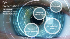
Transcript: Jasleen Brar Pavanpreet Brar Kavilen Valaydon Macular Degeneration How does normal vision work? How does normal vision works? -Normal vision works when light rays enter the eye through the cornea, which bends or refracts the light rays that pass through the pupil(which is a round hole ). -Then the light rays pass through the lens which changes shape and bend further and focus them on the retina. - which process the light rays into light impulses through millions of tiny nerve which are cells those cells are then sent to the optic nerve which then send image the head. Eye image Video Video Here are some more information about how the eye works. Macular Degeneration Macular Degeneration -Macular Degeneration is the cause of when people over 60 lose their vision. - It happens when the macula, which is the central area of the retina breaks down. There are two forms of macular degeneration, dry form and wet form. - The dry form of macular degeneration is when the appearance of yellow deposits called, drusen come into the macula. They don’t do much harm at first, but when they grow they can cause a person to have blind spots in their vision. - Next, the wet form of degeneration is when uncommon blood vessels come from underneath the macula and start to leak. This causes a person to see straight as wavy, or blind spots. picture of how Macular Degeneration looks Macular Degeneration Macular Degeneration -A way to cure macular degeneration is using anti-angiogenic drugs which is a medication a doctor injects to stop more blood vessels from leaking -Another cure to macular Degeneration is laser therapy, this can remove the blood vessels -Other things you can try are vitamins, vision aids or retinal translocation - Places you can go to get this cured is hospitals or clinics How is it Cured and where How It's Cured Jaundice (Yellow Eyes) Jaundice Jaundice is a yellowing of the whites of the eyes that happens when the body does not process properly. A waste material gets built up in the eye. This may be due to a problem in the liver. Symptoms include a yellow tinge to the skin and whites of the eyes, and itchiness. There are three types of Jaundice: Hepatocellular jaundice, Hemolytic jaundice,and Obstructive jaundice. Bibliography Bibliography https://www.medicalnewstoday.com>/articles/165749.phphttps://www.aoa.org/patients-and-public/resources-for-teachers/how-your-eyes-work https://www.webmd.com/eye-health/macular-degeneration/age-related-macular-degeneration-overview#1http://www.allaboutvision.com/conditions/amd.htm

Eye - Presentation

Transcript: OUR BUILT IN OPTICAL DEVICE THE EYE Elisabeth Kanczula The Eye OVERVIEW The eye is a complex organ found in animals. About 70% of sensory receptors are found in the eye. Its primary function is to detect light, and send signals up the optic nerve to be processed by the brain. The brain then forms this information into the image we see. ANATOMY OF THE EYE ANATOMY The human eye has multiple key components that work together to help form an image. The components of the eye that are most commonly talked about are the cornea, iris, lens, retina, pupil, vitreous gel, and optic nerve Cornea The Cornea Around the eye is a completely transparent, protective layer of cells called the cornea. It is about as thick as a credit card As well as being a protective layer, it also holds the eye together. The cornea also refracts the light rays into our eye, ensuring the light rays travel correct into our eye to produce an image. The Iris Iris The iris is a muscle that controls how much light is let into the eye. It dilates and opens when it is in dim light to take in as much light as possible. The iris also contracts in bright light, to prevent too much light from getting into our pupil. Interestingly enough, everyone's iris can be a different colour, depending on how much melanin is in your iris. This melanin is meant to keep the iris opaque and not allow light through. Lens Lens The lens is located between the pupil and the iris, and is formed by a transparent tissue. It focuses light that passes through the vitreous gel in our eye and to our retina, which helps produce a more accurate image. The lens in the human eye is a convex shape, thicker at the middle and smaller at the edges. Retina Retina When light arrives in through the pupil, at first, it is only wavelengths of light. These wavelengths are converted into nerve impulses that the brain can register, which allow us to see detail and the image as we do by our retina. There are two types of cells in the retina, rods and cones. They process different types of light. Rods process black and white, as well as contrast. Cone cells come in three colours, red, blue and green, and allow us to see detail and colour. The Pupil Pupil Looking the centre of the iris, you will see a black circle. This is your pupil! Along with the iris, the pupil controls the light that is let into your eye. The pupil specifically is the hole at which the light enters the eye, allowed it to travel through the lens and eventually to the retina Our pupils appear black because all the light in the tissue of the pupil is absorbed in different ways. Vitreous Humor (Gel) Vitreous Gel The vitreous humor, or vitreous gel, is located behind the lens. As the name suggests, it is a clear gel that fills the eye. Light passes through the vitreous humor before it reaches the retina. The vitreous humor protects the eye and helps it hold its sphere shape. As well, the gel helps ensure the retina is properly connected to the wall of the eye. The Optic Nerve Optic Nerve Once the light is brought into the eye and an image is formed, this image is converted into nerve impulses. These nerve impulses get sent through the optic nerve These nerve impulses are processed by our brain, which produce the image we see of the object we are looking at. How Does the Eye Work, Then? AS AN OPTICAL DEVICE The components of the eye all work together to create the final image we see. FUNCTION DETAILS Light reflects off of an object and is refracted by the cornea. The light then travels into our eye through the pupil. After this, the light rays are further refracted by the lens in our eye. This is so the light is focused on our retina. The image produced by the lens is smaller and inverted, opposed to the image we see. The retina, consisting of rod and cone cells, convert the light into electric impulses. These impulses are then sent up to the brain by the optic nerve. Once the brain receives these signals, the image we see is formed. COMMON VISION ISSUES COMMON VISION ISSUES While our eyes and intricate, complex, well running organs the majority of the time, this does not mean there can't be problems or tricks. Only 35 percent of the population of adults have full functioning vision. Errors can range, such as optical illusions, colorblindness, blurred vision, or near/farsightedness MYOPIA AND HYPEROPIA Hyperopia and Myopia There are many common issues to explain why a persons vision may appear blurry when they look at objects far away or up close. Two common ones are hyperopia, affecting about five to ten percent of the population, and myopia, affecting about thirty percent of the population. Hyperopia Hyperopia is the scientific name given for what we commonly know as farsightedness. Farsightedness occurs when the image produced in the eye is formed behind the retina, as opposed to on the retina. Hyperopia can occur due to the eyeball being too short or the cornea being too flat. HYPEROPIA Myopia MYOPIA Myopia is the medical term for

Eye Presentation

Transcript: In school Eye Presentation The optic nerve is affected with optic nerve hypoplasia, the nerve is underdeveloped and is present at birth. Reading off the boar, projector.ect Saying "do you see that right there' Looking for small objects, or even objects in general of not familiar. Saying "it's right over there Where my finger is pointing." other vision related phraes such as this. Diagnosis- Optic Nerve Hypoplasia Things that are not helpful Things that are helpful In my experience having Optic Nerve Hypoplasia, I can primary see most of what everything else see's, but I cannot see smaller objects, or things that fly or move quickly. For instance, I cannot see a cats claws retract, or I cannot see birds in the sky, or even on land.. With my visual acuity I see the world through one eye. I see evverything through my right eye, and a smaller percentage through my ledt. I do not see through both eyes at the same time, instead I see everything through one eye and small bits and peices through the other. When trying to see through both eyes, I can see colors, details, shadowing, and glare all through my right eye. In my left I can see basic structures of things, without a lot of detail. I can try to see only out of my left eye, but when I do I often get a temporary head ache. Wen seeing through my left eye only I can see details color, and shadows but ta lesser distance then m right. When seeing through both eyes I know that I can see details through my left eye, but I can not just see them, in a way I have to "pull them out" What is not helpful at school Simulation Sitting closer t the board Extra time on tests Printing out hard copies of what is on the board( depends) Analysis

Now you can make any subject more engaging and memorable