You're about to create your best presentation ever

Eeg Powerpoint Template

Create your presentation by reusing a template from our community or transition your PowerPoint deck into a visually compelling Prezi presentation.

EEG

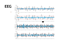
Transcript: b Y 2 X g m 1 p ELECTROENCEPHALOGRAM SAJAN MUHAMMED 6B EEG F An EEG is a test that detects abnormalities in your brain waves, or in the electrical activity of your brain E WHY? monitor alertness, coma and brain death; locate areas of damage following head injury, stroke, tumor, etc.; test afferent pathways (by evoked potentials); monitor cognitive engagement (alpha rhythm); W EEG ACQUISITION Electrode caps, conductive jelly, ruler, injection and aid for disinfection. EEG amplifier unit, PC/laptop In all modalities but EEG ,the sensors are heavy. EEG is the only modality that does not require the head/ body to be fixed. EEG might enable the monitoring of the brain functions of unconstrained participants performing normal tasks in the ELECTRODES ELECTRODES PLACEMENT P Preparation Food and medications Avoid anything with caffeine on the day of the test because it can affect the test results. Take your usual medications unless instructed otherwise. Other precautions Wash your hair the night before or the day of the test, but don't use conditioners, hair creams, sprays or styling gels. Hair products can make it harder for the sticky patches that hold the electrodes to adhere to your scalp. If you're supposed to sleep during your EEG test, your doctor might ask you to sleep less or avoid sleep the night before your test. P Procedure An EEG can be done in the doctor's office Lie on a bed Attach the electrode on the scalp Before attach to follow the preparation to get EEG Each electrodes are connected to an amplifier and record the EEG waves on recording machine EEG NORMAL EEG

EEG

Transcript: Electroencephlocardiograms used to measure effects http://www.webmd.com/epilepsy/electroencephalogram-eeg-21508 * 1980's- invention of computers allowed EEG brain topography to develop http://www.cerebromente.org.br/n03/tecnologia/historia.htm Future * The patient's head is covered in a gel to improve the conduction from the skull to the electrodes. It is important for the patients hair to be clean so there is little to no interference. * Many electrodes on placed on the patients skull, and a computer records the response. * The patient is usually sleep deprived and sometimes subject to flashing lights and hyperventilation. This is done because usually abnormalities are most apparent when the brain is under stress. Spectoral analysis 3-D brain reconstructions * There are very little risks because it measures brain waves on the surface of the scalp. * It can be riskier for people with seizure disorders because of the flashing lights and hyperventilating techniques used during this procedure, which can trigger a seizure. How is this procedure performed? Uses in medicine * 1875-Richard Canton discovers the EEG from the exposed brains of rabbits and monkies * Can detect whether or not a person is brain dead or just in a coma. * Effective in detecting and diagnosing the biological aspects of psychological and neurodegnerative disorders. * Treatements with an EEG can monitor the frequency of brain wave changes in children with ADHD and normalize them. * EEG biofeedback can be used improve attitude and emotional balance in people with learning disabilities, migrines, chronic pain, and teeth grinding. * Neurobiofeedback gives a person better control of biological and phsyiological functions. * EEG stimulation has been proven to improve memory and accelerate learning. Also, it improves sleeping patterns in people who have sleeping difficulties. Sources * Because EEG's measure action potentials (electic potentials), they are very similar to a voltmeter. Waves of ions reach the scalp, and affect the electrons on the metal electrodes. This difference is measured by the voltmeter and recorded over time. Risks involved in EEG tests *Advancement of computers has led to many great things http://bml.ym.edu.tw/ibs/brain/curricurium/952curricurium/file/MEG_EEG_Clinical%20applications.pdf * Because it measure electrical activity, it is not suggested for recording all types of brain activity. It is usually used to detect: http://www.nlm.nih.gov/medlineplus/ency/article/003931.htm http://benefitof.net/benefits-of-eeg/ http://www.edonline.com/collegecompass/oohb0114.htm http://www.medicalengineer.co.uk/pages/medical-imaging/eeg-applications-in-modern-medicine.html Physics Principles Electrode patch wires are placed on designated spots on the head. These electrods record four types of brainwaves according to frequencies: alpha, beta, theta, and delta. EEG technicians study the recorded changes in the brain impulses intently and then proceed with diagnosing the patient. A single axon potential is too small to be picked up, so EEG's actually measure the summation of thousands or millions of neurons synchronously firing. Who is this test for? History History *1957- W Gray Walter invented the toposcope- a series of cathode ray tubes attached to electrodes attached to the skull. Biofeedback (neurofeedback) can be used to diagnose and treat patients. Biofeedback is the information provided from an EEG. EEG's from a physics perspective How it works * Tumors * Focal disease epilepsy arterivenous mal-formations stroke * Disturbances in consciousness and viligance narcolepsy coma * Effects of withdrawal from psychoactive drugs * Effects of infectious diseases Braim Meningites * Identifying a psychological disorder with a physical cause schizophrenia dementias hyperactivity depression brain atrophy attention deficit disorders Used any time the electical potential in the brain needs to be monitered http://en.wikipedia.org/wiki/Electroencephalography Improvment of life * 1929- Hans Berger measured the human brains electrical activity on the scalp

EEG

Transcript: Classification SCHEME for EEG based Motor Activities inducing ICA using Machine Learning Presented By: Name: Pranali Kokate ID: 2019PEB5431 Supervisor: Dr. Amit Joshi Introduction Neurologists are increasingly developing methods and means to integrate brains and machines. The most significant of them are brain-computer interfaces (BCIs), which can provide alternative modes of communication and enables control of external equipment even in severe situations of impairment. In this field, research is developing in all aspects, be it the large scope of EEG-based BCI[2] controls, the technological depth involved, and its usability for impaired people and the rest of the population. Every day, new and creative scientific advancements are being investigated in order to improve the lives of millions of people all over the world. Many researchers have developed EEG-based BCI systems to overcome the problem. An Electroencephalogram[1] is created by collecting and storing electrical signals taken from the brain. Thus providing an ability to extract relevant information about brain activities. Introduction [1]Guangyi Zhang, Ali Etemad, A.S.Moghaddam, Y Zhang, "Classification of Hand Movements from EEG Using a Deep Attention based LSTM Network." IEEE Senors Journal ,March 15 2020. [2] S. N. Abdulkader, A. Atia, and M.-S. M. Mostafa, “Brain computer interfacing: Applications and challenges,” Egyptian Inform. J., vol. 16,pp. 213–230, Jul. 2015 Techniques in bcI Why EEg ? There are different techniques in BCI Invasive as well as Non-invasive [3] Magnetoencephalography (MEG) : MEG is a noninvasive neurological recording technique that captures the magnetic activities produced by natural sources like the electrical impulses of neurons in the brain. Functional Magnetic Resonance Imaging: fMRI is yet another non-invasive technique of brain scanning method that uses electromagnetic fields to detect and record. Functional Near-Infrared Spectroscopy (fNIRS): The fNIRS is a noninvasive brain wave collection method that uses near-infrared light to quantify alterations in localised cerebral metabolism while performing particular neural activity. EEG based BCI standsout from its conterparts due its long-lasting usability, high temporal resolution, ease of installation, and low prices. Sanei, S 2013, Adaptive Processing of Brain Signals, John Wiley & Sons, Incorporated, Hoboken. Available from: ProQuest Ebook Central. State-of-art technique adopted with different EEG parameters literature Survey We reviewed various available data sets related to motor imagery and execution activities. Several features associated with EEG signals. Different Pre-processing technique. Various classification algorithms from machine learning to deep learning. Research gap Research gap Proper selection of channels[1]. Despite applying the digital filter to EEG, contaminated data still exists, because such data is generated by eye blinks and head movements. To obtain cleaned EEG data, we removed the contamination factors by an independent component analysis (ICA) which is commonly used to decompose the brain signals into statistically independent components (ICs). [3] The fundamental disadvantage of EEG signals is that they are vulnerable to both external and other biological influences. There must be a thorough discussion as well as some conventional ways for removing the tainted noises. Validation of the applied procedures is required. [3] Brain-Controlled Robotic Arm System Based on Multi-Directional CNN-BiLSTM Network Using EEG Signals, Ji-Hoon Jeong , Kyung-Hwan Shim , Dong-Joo Kim , and Seong-Whan Lee , 5, May 2020 Objective 1.The identification and extraction of relevant signals from the raw EEG data is likely the primary goal for that we considered a publicly available dataset. 2. Understanding the structure of the brain for effective electrode selection. 3. To implement an algorithm for removing artifacts and cleaning signals. 4. To propose a classification Machine Learning model using ANN. 5. To develop a deep learning-based framework for an EEG-based BCI system. Methodology Methodology data-set BCI Dataset for motor imagery and motor execution The EEG Movement Dataset was used in this study. The dataset includes 109 subjects and has been collected using a BCI 2000 system. [7] Participants were asked to perform three actions: rest (T0), left hand movement (T1), and right hand movement (T2). Each experiment consisted of 15 iterations, where T0 was followed by a visual stimulus, randomly selecting either T1 or T2. This 15-pair movement process was repeated 3 times. The data set consists of 109 subjects and each subject is performing 14 tasks among 14 tasks we considered only 6 tasks. Here 3,7 and 11 runs represent motor execution class while 4,8 and 12 runs represent motor imagery class. The dataset contains 64-channels of EEG, recorded at a sampling frequency of 160 Hz. Figure illustrates a sample EEG recording and the three actions T0,T1, and T2. [7]

Now you can make any subject more engaging and memorable